A Guide to 9103 Picoammeter Compatibility

Print Friendly, PDF & Email

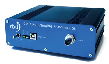
RBD’s 9103 USB Picoammeter and Actuel application (included in your purchase) are compatible with a number of hardware devices and operating systems. The complex nature of operating systems and hardware, as well as the differences between 9103 hardware and software compatibility, render a simple compatibility table somewhat lacking in clarity. This guide will hopefully serve to answer the most common questions concerning 9103 Picoammeter compatibility.

9103 Picoammeter

9103 Picoammeter

Using the 9103 Picoammeter with Either Actuel or a Custom Application

While many customers will only ever need to use the 9103 with the Actuel data-logging / graphing software, others will want custom or advanced capability and compatibility. Happily, the 9103 is fully programmable in a simple, well-documented ASCII language. We’ll post more in-depth coverage of 9103 programming in the near future. For now, just keep in mind that writing an application to communicate with the 9103 is relatively simple, though of course your application can be as complex as you want it to be.

9103 Hardware / Firmware Compatibility

The 9103 uses industry-standard USB interface hardware based on a chip from FTDI. That means the 9103 is compatible with the all the operating systems supported by FTDI, as long as the device has a compliant USB port. If you are planning to program your own custom control/display application for the 9103, the following operating systems are supported:

Windows 8 x64
Windows 7
Windows 7 x64
Windows Server 2008
Windows Server 2008 x64
Windows Vista
Windows Vista x64
Windows Server 2003
Windows Server 2003 x64
Windows XP
Windows XP x64
Windows ME
Windows 98
Linux
Mac OS X
Mac OS 9
Mac OS 8
Windows CE.NET (Version 4.2 and greater)
Android

Note that the the iOS operating system, installed on the iPhone, iPad, and iPod devices, is not currently supported.

More information on FTDI’s operating system support, as well as drivers for those operating systems, can be found here:

http://www.ftdichip.com/FTDrivers.htm

9103 Actuel Application Compatibility

The Actuel application provided with the 9103 is a Windows PC-only application and compatible with the following operating systems.

Windows 8 x64
Windows 7
Windows 7 x64
Windows Vista
Windows Vista x64
Windows XP (Service Pack 3)
Windows XP x64 (Service Pack 3)

Note that the Microsoft .NET 3.5 library, included with the Actuel application and freely available from Microsoft, must also be installed.

Special Considerations for Using the 9103 on Apple Mac Hardware

While the Actuel application is a Windows application, keep in mind that Apple Mac line now runs on Intel hardware, meaning that Actuel is compatible with any Mac installation running Windows. In fact, Actuel was developed on a MacBook Pro running Windows as a virtual operating system.

Also, since USB port compatibility is operating-system dependent, you can (and must, in fact), use the drivers compatible with your operating system regardless of whether you are running PC or Mac hardware when you creating a custom application for your 9103. If, for example, you are running Linux on a Mac (using Bootcamp), you would use the FTDI drivers for Linux.

The 9103 hardware is compatible with all the operating systems listed for the FTDI drivers when that OS is running on Mac hardware.

Actuel is compatible with the Windows operating systems listed above when running on Mac hardware. The following configurations are examples:

Bootcamp (runs Windows as a boot drive independent from iOS)
VMWare Fusion (runs as a virtual operating system on iOS)
Parallels (similar to Fusion; untested as of this posting)

National Instruments LabView Compatibility

LabView provides a rich environment for programming a current measurement device such as the 9103 USB Picoammeter, which is compatible for any operating system and hardware device that is supported by BOTH Labview and FTDI.

Operating systems supported by LabView can be found here:
http://www.ni.com/labview/os-support/

While RBD does not directly provide LabView instrument drivers/scripts or programming support, a third-party “starter” kit is freely available from our 9103 downloads page:
https://www.rbdinstruments.com/Products/Picoammeter/Downloads.html

I hope this answers most of your 9103 compatibility questions. Keep an eye out for another blog post in the near future with more helpful information on programming the 9103 using simple ASCII commands.


How to test an ion gauge filament

Print Friendly, PDF & Email

This post will explain how to test and replace the nude ion gauge filament assembly on a Physical Electronic (PHI) X-ray photoelectron, Auger electron or SIMS system. Look at the pictures at the bottom of the post before you read the procedures.

Background – On most PHI surface analysis systems the ion gauge filament is located either above the table tops in back of the vacuum chamber, or just under the tabletops.  The newer (as in less than 30 years old) systems have a cover that protects the user from the exposed electrical connections to the ion gauge pins. On the oldest PHI systems the ion gauge pins are exposed, but located under the table tops and difficult to access (and so relatively safe).

Here are links to some videos that explain how an  ion gauge works –

https://www.youtube.com/watch?v=IKKuWeEShM4

https://www.youtube.com/watch?v=6zv_Y0_vwsg

How to measure the resistance on the ion gauge:

  1. Turn off the DGCIII (or other brand of) ion gauge control.  This is not only the first step; it is the most important step! Ion gauge controls such as the DGCIII used on older PHI systems have about 200 volts of DC on the grid. If you do not turn off the DGCIII (or other) ion gauge control before removing the wires to the ion gauge you will likely receive a potentially lethal electrical shock. If you are not familiar with working safely with electricity then refer this procedure to qualified personnel.  Or, turn off the DGCIII and also and also unplug the 120 VAC power cord on the back of the DGCIII and then there is no danger of electrical shock.
  2. Loosen the set screws on the shield retaining collar. Do not loosen or remove the bolts that connect the ion gauge to the system! See the pictures at the bottom of this post for clarification.
  3. Loosen the strain relief screws and slide the shield out and away from the ion gauge, being careful to support the wires.
  4. Using an 048-4 spline wrench, loosen the ion gauge coupler set screws by turning the set screws closest to the flange CCW 1 to 2 turns and then gently pulling the coupler and wires off of the ion gauge pins. TIP: As you remove the couplers turn the set screws CW 1 turn so that they do not fall out of the couplers. RBD provides the 048-4 spline wrench and the setscrews.
  5. Use an ohmmeter and measure the resistance between the center filament pin (common) to the outside two filament pins. See the picture below. The pins resemble a smiley face and the filaments are the smile. The grid is the eyes (some ion gauges have 2 grid pins, some only one), and the collector is the center pin (nose). The filament resistances should be 1 ohm or less when measured from the center filament post to the outside two filament post. If a filament is burnt out (open) then the resistance will be infinite or some high value if there is a tungsten coating on the filament base.
  6. If one filament is burnt out but the other one is good, then you can switch filaments.  If you have 3 wires connected to the filaments then swap the outside two filament connectors. If you have just two filament wires, then move the outside filament wire to the other side.
  7. If both filaments are open, then the filaments need to be replaced. See the replacement procedure in the following section.

ion-gauge-wire-connection-types

ion-gauge-wire-connectionsHow to replace the ion gauge filaments:

  1. Vent the chamber.
  2. If not already done, remove the connectors from the ion gauge as per the previous procedure.
  3. Remove the bolts from the ion gauge flange.
  4. Remove the shield retaining collar.
  5. Carefully remove the ion gauge.
  6. Loosen the top set screws on the 3 filament base connectors. These are typically .050 hex screws.
  7. Remove the old filament assembly.
  8. Install the new filament assembly and tighten the set screws. Make sure that the filaments are parallel with the grid.
  9. Use a new copper gasket and place the ion gauge up to the flange. Make sure that the filaments are facing down. They will not line up perfectly parallel, so just choose the best position where the bolt holes line up. By facing the filaments down you will prevent any debris from falling onto the grid which may short out and damage the ion gauge control.
  10. Place the shield retaining ring up next to the ion gauge flange and rotate it so that the set screws in the shield retaining ring are accessible.
  11. Insert the bolts and tighten the flange.
  12. Reattach the ion gauge couplers. Make sure that the pins are bent slightly in towards the center collector wire so that none of the pins will short to the shield when it is installed.
  13. Carefully slide the shield over the wires and press the shield firmly into the shield retaining collar.
  14. Tighten the shield retaining set screws.
  15. Slightly tighten the strain relief screws.

That’s it!  Pump the system down and the ion gauge is ready to turn on once you get into the 10-4 Torr or better vacuum.

RBD Instruments provides replacement filament assemblies, complete ion gauge assemblies and the required spline and Allen wrenches. Contact us for more information.

This slideshow requires JavaScript.

AugerScan AES XPS software Tutorial

Print Friendly, PDF & Email

AugerScan is RBD Instrument’s widely used software program which acquires and massages data on older Physical Electronics PHI X-ray photoelectron and AES Auger surface analysis systems and components. AugerScan is used in conjunction with the RBD Instruments 147 PC Interface unit. The 147 connects to the PC and provides a hardware interface between the software and the system hardware.

This post will explain all of the features and settings in AugerScan and how to configure the software for your particular system type.

augerscan-pulldown-menus

Beginning with the first pull down at the top of the status bar we have the File menu.

New New acquisition. Acquisition types include Alignment, survey, multiplex and depth profiles. You can also select the technique – XPS, AES or SIMS.
Open Open a previously acquired or saved file. You can save a file without acquiring it, in which case you can use it as a template for acquisitions of common settings.  For example, you may want to make a file called Elastic Peak which would be used to set the sample to analyzer distance when acquiring AES data.
Close Standard Windows command
Save Standard Windows command
Save as Standard Windows command
Import Imports AugerScan 1.X versions into the 3.X format
Export as Text Export file to ASCII format.  The ASCII format can be used for programs such as Excel and is also the format used by CASAXPS to read in AugerScan files.
Export All as Text Exports multiple files to ASCII format.
Export to Quases Export file to Quases formatQuases software download
Export all to Quases Export selected files to Quases format
Export to Multipak Export to older versions of the PHI Multipak format. Most new surface analysis systems use (or offer as an option) CASAXPS for advanced data massage functions.
Print Standard Windows command
Print Preview Standard Windows command
Print Setup Standard Windows command
Page Setup Standard Windows command
Create Reserved for future use, this command was intended for creating a new Windows object. However, Windows has changed so much that this feature is no longer relevant.
Recent Files A list of the most recent files that were opened. Currently this lists the last 4 files.
Exit Exits and closes AugerScan

The next pull down on the status bar is the Edit menu.

Copy Copies the open file to the Windows clip board. This is used to copy and then paste a survey into Word or some other windows program.
Copy as ASCII Copies the data in an ASCII format to the Windows clipboard.  This feature is used to copy and then paste data in Excel or some other windows program.
Paste Pastes the clipboard into another Windows program
Delete Not used
Mark/Track This is a cursor that displays the intersected energy in eV and the counts in the status bar. It is also used to select elements for the atomic concentration calculation for AES data.
Markers Displays the selected markers or endpoints. When selected the element marker will be displayed on the displayed data peak or in table form. When checked, the selection will be included in the atomic concentration calculation.
Select Endpoints Allows the user to select the endpoints used for the atomic concentration calculation. To use – move the cursor to the desired left end point. Press and hold the left mouse button. Drag the cursor to the desired right hand endpoint, release left mouse button.  This feature is helpful for selecting the peak of interest.
Endpoints Displays the selected endpoints
Annotation Standard Windows command
Element Table Tables used for atomic concentration calculation for AES and XPS, for markers only in SIMS. AES table includes sensitivity factors for 3, 5 and 10kV beam voltage. XPS sensitivity factors include peak and area modes for standard 15kV dual anode and monochromator X-ray sources.
Insert New Object Standard Windows command – seldom used command for inserting things like table or pictures into the data display.
Links Reserved for future use.
Object Reserved for future use.

The next pull down on the status bar is the View menu.

Toolbar The toolbars contain Icon shortcuts to commands and dialog boxes. Once you know these shortcuts they are easier to use than the pull down menus. The View Toolbar command allows the user to enable or disable the toolbars:

  • Main
  • Acquisition
  • Region Selection

We recommend that the tool bars are always selected.

Status bar This is the little box at the bottom of the Augerscan window. It displays pertinent information such as the cursor values and the status of data acquisitions.
Zoom The zoom feature allows you to view an area of interest in the data. To use the Zoom feature, move the magnifying glass cursor to the upper left hand corner of the area of interest. Click the left hand mouse button and drag the box to the lower right hand limit of the area that you want to view. Release the left mouse button. You can perform multiple zooms in.
Restore Un-zooms to the original data display.
Raw Data Displays the raw (original) data.
Transformed data Displays the massaged (transformed) data.
Cycles For depth profiles All region selection, selects cycles as the X axis on the displayed data.
Time Displays the depth profile X axis as time.
Depth Displays the depth profile X axis as depth. This is calculated from the sputter rate set in the Electron/Ion gun properties dialog box.
Expand X axis Expands the X axis display to allow room for text or an image.
Options Allows the user to set options for:

  • General
  • Line Style
  • Annotations
  • Fonts
  • Curve fitting
  • Graph

The next pull down on the status bar is the Acquisition menu.

Start Starts the selected acquisition
Stop Stops the acquisition at the end of the next sweep or cycle
Stop Now Stops the acquisition immediately and the data is not saved
Continue Continues the selected acquisition. This is useful if you want to stop a depth profile at an interface and then acquire a survey, then continue the profile.
Settings Displays all of the settings for the selected acquisition.
Edit Allows the user to remove regions or cycles from a depth profile, and edit the lower and upper limits in surveys.
Sample Properties This menu is where user can enter comments for samples. Typically this is used for 8 sample specimen stages on XPS systems.
New Survey Wide range scan, typically 30 to 1030 or 2030eV for AES and 1100 to 0 binding energy for XPS.
New Multiplex Series of narrow surveys, allows the user to acquire data only on the elements of interest.
New Depth Profile A series of multiplexes with combined with sputter etching acquires the elements of interest vs. depth.
New Alignment (peak) Used to set the elastic peak in AES, and to optimize the position of the sample or X-ray source in XPS.  Data is thrown out at the end of each sweep.
New 3 point depth profile For AES only, a 3 point depth profile makes it possible to get more elements into a depth profile than is practical with the standard depth profile. Since there are only 3 points selected per region (rather than the typical 20 to 50 points per region in a standard depth profile) you can get more elements into the same amount of acquisition time. Good for general trends only as a 3 point profile cannot use the atomic concentration calculation.
Technique Select the technique – AES ( Auger Electron Spectroscopy), XPS (X-ray photoelectron spectroscopy) or SIMS (Secondary Ion Mass Spectroscopy)
Options Allows the user to set options related to acquisitions. The most commonly used is the Prompt to Save after acquiring.

The next pull down on the status bar is the Data menu.

Revert to Raw Data Removes all previous data massage functions and restores the display to the original data.
Edit Raw Data A text editor, this command allows the user to edit out data spikes
Differentiate Normalizes background and displays data in peak to peak form.
Smooth Savitzky and Golay smoothing algorithm.
Convert to Counts Data in Augerscan is displayed in counts per second. This command converts the counts per second value to actual counts.
Shift Shifts the data up or down as specified by the use. Useful for compensating for small amounts of sample charging.
Depth Profile Smooth Smoothes Depth profile display. Similar to the data smooth command.
Deconvolute X-ray Line Fourier transform function that sharpens the XPS peaks
Satellite Subtraction Removes satellite peaks from data which improves the resolution (FWHM).
Baseline Removes the background from the selected endpoints.
Curve Fit Used for fitting hypothetical (possible) peaks into existing multiplex data.
Delete Curve Fit Deletes selected curve fit
Atomic Concentration Performs atomic concentration calculation. Peaks and endpoints need to be selected first. If the sensitivity factors are not in the element table you will need to add them before performing this function.
SIMS Relative Sensitivity Factor Used for SIMS data analysis
SIMS Atomic Density Used for SIMS data analysis
Options Set the number of points for smooth and differentiate, list the range for markers and select peak or area for XPS atomic concentrations.

The next pull down on the status bar is the System menu.

Hardware Properties Selects hardware properties for your system type. This is where you tell AugerScan the specific controls that your system has. Depending on the vintage and type of your system, most model numbers are located in the upper left hand corner of the electronic unit.  For card rack type systems, the model numbers are listed on the bottom of the mother board for each board set in the back of the electronics bay. You also set the input (detector), X-ray anode type and energy, and can calibrate the peak positions (scale factor). We provide calibration information with each 147 upgrade.
Multiplier Properties Sets the multiplier voltage for each technique. For AES, the multiplier can be set automatically for different electron beam currents by selecting the appropriate V/F preamp. Select the Preamp Defaults after first setting your preamp type (typically 96A/B) in the Hardware Properties dialog box.
Gun Properties For booking purposes only, this is where the user enters the typical settings for the electron and ion gun. The sputter rate value is used for the atomic concentration depth view.
Diagnostics Used to test the RBD 147 PC interface unit, for most users the DR11 test and DR11 data entry sections are useful for occasional troubleshooting or calibration of electronic components. To run the full diagnostics special jumper cables are needed.
Sputter Simple sputter timer is useful for sputter cleaning samples before analysis.
Ion Gun Control Opens up the ion gun control dialog box for digital ion gun controls such as the 77-070 (04-300 ion gun), 20-520 (Duoplasmatron ion gun) and 11-066/68 (04-303 and 06-350 ion guns).
Stage Control Controls the movement of digitally controlled specimen stages
Neutralizer Control Controls the 04-085 and 04-090 neutralizer
SIMS Positional Alignment Used for mechanically adjusting the SIMS optics to the position of the ion beam.
XPS Mapping Used for 5600, 5700 and 5800 XPS system that have the mapping hardware option. Due to the small count rate at the smallest aperture size this is not really a practical feature. New state of the art imaging XPS systems have much higher count rates while imaging and much better spatial resolution.
Automation Used primarily for digitally controlled specimen stages, this dialog box allows the used to stack up a number of commands and then run all of the commands as a batch.
Enable Batch Feedback Control No longer used.
Disable Batch Feedback control No longer used.

The next pull down on the status bar is the Window menu.

New Window Standard Windows command
Cascade Standard Windows command
Tile Horizontally Standard Windows command
Tile Vertically Standard Windows command
Arrange Icons Standard Windows command
Display All Regions Opens a window for each region in a multiplex or depth profile.  Currently you need to select this option after the acquisition starts or is completed. The next release of AugerScan will have an option for this to happen automatically.
Recent Acquisitions Lists the 4 most recent files.

The next pull down on the status bar is the Help menu.

Help

The original Help system (largely unchanged since Windows 3.1) was phased out in Windows Vista. The context-sensitive help – also known as “what’s this?” or “right-click” help cannot be replaced (this was the information you would typically see for each field in a dialog box, for example). However, for Windows Vista through 8.1, Microsoft does provide separate downloads for the legacy help system; it’s just no longer installed in the operating system.

You can find most of those files on this Microsoft support page (scroll down to “Resolution”) For security reasons, Microsoft no longer supports this help format at all in Windows 10, and there are no third-party solutions available.

However, we’ve translated most of RBD’s help to HTML for both AugerScan and AugerMap. In each case, simply  unzip the file to a convenient folder and run “index.html” in your browser.

augerscan-iconsThe images below list the commands for the shortcut Icons on the Main and Acquisition tool-bars. Once you learn these commands they make it very simple and fast to acquire and massage data.

Augerscan-Icons-vert

Augerscan Shortcut Icons