Homemade Titanium Sublimation Pump

Print Friendly, PDF & Email

In this post I show how we made a small homemade titanium sublimation pump for an 8” Kimball Physics spherical octagon UHV vacuum chamber.

Our little chamber has a 60 l/s ion pump, but even with baking (both IR and UVC), we were able to get only into the low 10-8 to high 10-9 Torr range.  However, using the little titanium sublimation pump, which we “Frankensteined” together using parts we had readily available, allowed us to get in the low 10-9 to high 10-10 Torr range, a factor of ten improvement.

A titanium sublimation pump works by heating a titanium filament wire to about 1300 degrees C. That is hot enough to create titanium gas molecules (sublimate) but not so hot that the filament wire melts.  The sublimated titanium deposits on the wall of the chamber (or preferably on a shield wall) and forms a thin film. This layer of titanium is very reactive and will bond with other molecules in the vacuum chamber such as CO and O2. Disassociated hydrogen and water vapor also diffuse into the titanium layer.

The reactivity of the titanium film is increased with lower temperatures, but most titanium sublimation pumps are operated at room temperature. Over time, the titanium film will become coated and need to be replenished. All commercial titanium sublimation pumps have 3 to 4 filaments so that when one filament burns out you can switch to another. Those filaments are also relatively thick in diameter at 12 gauge (.080”) and need about 50 amps of current to operate.

For our homemade titanium sublimation pump, we used 24 gauge (.020”) so that we could operate at a much lower current of 4 amps.  We also only have one filament.

Before I show how we made our homemade titanium sublimation pump, here are links to some videos on how TSPs work:

https://www.youtube.com/watch?v=j5Y7m2ZJfgg

https://www.youtube.com/watch?v=9vJedaxRsxI

The first thing that we needed was a 2-pin electrical feedthrough on a 2.75” CF flange.  For that we used a Getter pump flange from a PHI 04-303 ion gun as shown below.

filament_wire_connections

filament_wire_connections

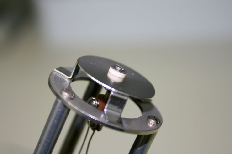
We then needed to somehow support and electrically isolate the TSP filament wire. To do that we used a coupler and some little shoulder washers.

Ceramic_support

Ceramic_support

The top part of the getter pump assembly is conveniently designed to allow gas molecules to pass through but also block direct deposition of titanium into the vacuum chamber.

blocks_direct_deposition

Next we added a few turns into the titanium wire so that it would have a little bit of a spring to it. Then we connected the wire to the flange and support assembly. We have only one filament and so by effectively doubling the length of the wire we could also double the amount of titanium that we would be sublimating.

filament_wire

filament_wire

For a chamber wall we used a 2.75” nipple that has a tube ID of 1.6” and a length of 4”.  The larger the surface area the better, but for the size chamber that we have, we are limited to a small 2.75” nipple. We mounted this nipple on our chamber horizontally so that any flakes that form will not get into the chamber or ion pump.

2.75_inch_CF_nipple

2.75_inch_CF_nipple

For a power supply, we used a 30 volt 5 amp Lavolta.

30V_5amp_power_supply

30V_5amp_power_supply

After installing our homemade titanium sublimation pump into the chamber we pumped down and were ready to operate the TSP.

To operate our titanium sublimation pump, we slowly increased the power supply current while observing the color of the light coming off the TSP filament and also monitoring the chamber pressure.  The filament needs to be orange for the titanium to sublimate. Too hot and the lifetime of the filament will be reduced. Too low and the pumping effect is reduced. By experimenting we determined that about 3.8 amps DC was the correct amount of current.  Once that was determined, we could just periodically turn the TSP on for about 2 minutes at a time.  We did that 3 times over a 6-hour period and then let the chamber pump overnight. The next morning we were in the high 10-10 Torr range.  Success!

tsp_filament_in_operation

tsp_filament_in_operation

Conclusions:

  1. It is possible to make a small titanium sublimation pump using off-the-shelf components that will operate with less than 5 amps of DC current.
  2. Adding a titanium sublimation pump to a small chamber can help to get from HV to UHV.


Loading PHI specimen mounts

Print Friendly, PDF & Email

Loading PHI specimen mounts

This post explains the mechanics involved with loading PHI specimen mounts used on most of the older to current Physical Electronics surface analysis systems.

There are a number of different sizes and shapes of specimen (sample) mounts and they all use the same basic mounting and docking scheme.

The most common sample mount used on older Physical Electronics XPS and AES surface analysis systems is the Model 190 flat one inch diameter specimen holder shown below.

Model 190 sample mount

Model 190 sample mount

The basic concept is that there are two grooves on the outside of the sample mounts which are used to transfer the sample mount into the vacuum chamber, and to dock the sample mount to the specimen stage. The transfer arm has a fork on the end which connects to the bottom groove on the specimen mount and the specimen stage has three clips which snap into the bottom groove and hold the specimen mount to the stage.   The top groove is used to hold the specimen mount while mounting it to the transfer arm fork or in the case of scanning auger systems, to transfer to the parking attachment.

one_inch_sample_mount_from_side

Grooves on 190 specimen mount

Clips on specimen stage

Clips on specimen stage

There are two essential steps required to successfully dock and removing the specimen holders;

  1. Make sure that the specimen stage is perfectly centered under the transfer arm fork.
  2. The specimen stage center clip needs to be lined up with the notch on the transfer arm fork.
Line up intro fork notch to clip

Line up intro fork notch to clip

line up sample over specimen stage

line up sample over specimen stage

Once you have the location of the specimen stage micrometers for the load and un-load positions you can make some marks on the specimen stage micrometers so that you will be able to easily re-position the specimen stage for loading and unloading.

 Loading PHI sample mounts procedure:

  1. After you attach the sample that you want to analyze to the specimen holder (using screws and clips or carbon/silver tape). Back-fill the intro with dry nitrogen and mount the specimen holder to the sample fork. There are 2 different types of specimen mount holding tools as shown below. Or you can use a clean needle nose pliers. Pull the transfer arm out a little bit so that you can lower the specimen mount into the load lock and then move the transfer arm back in so that the fork slides over the bottom groove on the specimen mount.
    specimen mount  holding tool

    specimen mount holding tool

    sample holding tool

    sample holding tool

  2. Pump down the load lock by pressing the Pump Intro button on the AVC remote box.
  3. Move the specimen stage micrometers to the load sample position. The X and Y axis will be centered under the intro fork but the Z axis will be lowered from the dock position by at least 1 cm.
  4. After the load lock has pumped down (5 bars on the AVC remote plus a few more minutes – the longer you pump the sample, the lower the pressure burst and the less out-gassing you will have) press the  Intro Sample button on the AVC remote and the V1 gate valve will open.
  5. Move the intro arm forward to bring the sample into the vacuum chamber and push it all the way in until it hits the stop. If adjusted properly, the stop will be the correct position for docking the specimen mount.
  6. The specimen mount should be directly above the specimen stage clips.  If not adjust the X and Y on the specimen stage as needed.

    one_inch_sample_puck_above_specimen_stage

    one_inch_sample_puck_above_specimen_stage

  7. Raise the specimen stage Z axis so that the clips slide into the specimen holder bottom groove. You may need to slightly adjust the X Y or rotation (if so equipped) so that the specimen holder snaps into the clips.   Slightly moving the specimen stage rotation or the tilt on the transfer arm can also help the specimen mount to snap down into the clips on the specimen stage as you move the specimen stage Z axis.   If the specimen mount does not snap down with a minimal amount of force, back off and recheck the specimen stage X and Y position.  You do not want to damage any of the three clips on the specimen stage by crushing them. Once the clips have snapped in adjust the Z slightly until there is no gap between the bottom of the specimen holder and the specimen stage.

    one_inch_sample_puck_docking

    one_inch_sample_puck_docking

  8. After the specimen mount is docked to the specimen stage, slowly retract the intro fork until it separates from the specimen holder and then once clear, pull the intro arm all the way out of the chamber. The V1 gate valve will close automatically.

To remove a sample from the system, repeat the above procedure with the exception that the Z axis should be in the dock position so that the intro arm fork mounts to the lower groove of the specimen mount. Once you dock the load lock fork to the specimen mount, drop the Z axis on the specimen stage in order to separate the specimen holder from the specimen stage clips.

When the specimen holder is clear of the specimen stage you can pull the intro arm all the way out of the chamber and V1 will close automatically.

RBD Instruments provides new and used PHI specimen mounts.  We also provide the sample clip assembly. Contact us for more information.

RBD specimen mount part numbers

RBD specimen mount part numbers

This slideshow requires JavaScript.

X-ray source arcing

Print Friendly, PDF & Email

X-ray source arcing results in unstable XPS data and can also damage the x-ray source power supply or high voltage control.

Typically X-ray source arcing is caused by contamination on the anode, a coated anode support ceramic (the football ceramic) or a loose filament.

However one unseen cause of x-ray source arcing is when the high voltage cable to the x-ray source is not tightened all the way down until it snaps in. The procedure is simple – line up the slot on the cable with the two little tabs on the source connector. Then press down all the way and turn the cable collar clockwise until it snaps in.

On the newer 04-548 15kV dual anode sources this is easy to do as the connector is exposed. But the connector is recessed on the older dual anode sources and all of the mono sources.

To properly connect the high voltage cable on the older dual X-ray sources or a mono source you need to remove the cover on the source so that you can see the connector and make sure that the cable slots line up and that the cable locks down when the collar is turned fully clockwise.

high-voltage-connector-slots

high-voltage-connector-slots

high-voltage-connector-snapped-in

high-voltage-connector-snapped-in

If the high voltage cable is not snapped all the way down then it can arc at higher voltages and damage the cable connector, the source connector, or more typically both connectors.

The pictures below show damaged cable and source high voltage connectors.

melted x-ray source connector

melted x-ray source connector


burnt x-ray source connector

burnt x-ray source connector


Once arcing damage occurs the cable end and or source connector need to be replaced.

RBD Instruments provides the source connector and we can also repair or exchange your cable. Visit our website and look under Parts – Optics – X-ray Photoelectron Spectroscopy Model 04-500 and04-548 X-ray source parts or call us at 541 550 5016

Revised outgas procedure for PHI dual anode x-ray sources and single anode mono sources.

Out-gassing the filaments and conditioning the anode are essential steps needed to remove adsorbed gases from the filament area of any PHI X-ray source.

Recently I have seen a couple of instances where a 10-610 monochromator source was not properly out-gassed and the result was a contaminated anode and very low counts. So degassing the anode is essential for proper operation.

To prevent anode contamination, the anode needs to be degassed per the PHI manual. However I have found that by changing the order of the out-gas procedure steps that the amount of time it takes to out gas the source to full power can be significantly reduced.

The manual states that the out-gas procedure sequence is as follows:

  1. Outgas the filaments
  2. Condition the high voltage
  3. Degas the anode

But from a practical standpoint it makes more sense to degas the anode before conditioning the high voltage. The reason is that a degassed anode is less likely to arc.

So the faster way to out-gas an X-ray source is:

  1. Outgas the filaments
  2. Degas the anode
  3. Condition the high voltage


Step 1. Outgas the filaments.

You need to out-gas the filaments after new filaments have been installed or anytime the system has been brought up to air and baked out. For this procedure it is assumed that the system has been baked out. (The only bake out exception is if you have just replaced the 04-303 ion gun ionizer and back-filled the chamber with dry nitrogen).

  1. Turn on the 32-095/096 power.
  2. On the 32-095/6, press the Blue Out/Act out-gas activate button.
  3. Select both filaments
  4. Select the Mg filament (or filament 1)
  5. Slowly increase the amps to 3.5
  6. Select the Al filament (or filament 2)
  7. Slowly increase the amps to 3.5
  8. Let the filaments sit there for a few minutes and then slowly increase each filament to 4.5 amps.
  9. Let the filaments sit at 4.5 amps for a minimum of 4 hours (overnight is best).
  10. After out-gassing for at least 4 hours set the filament current to zero on both filaments and turn off the Out/Act out-gas button by pressing it one more time.

Step 2 Degas the Anode

  1. Set the beam voltage to 500V and turn it on.
  2. On the 32-095/6, press the Blue Out/Act out-gas activate button
  3. Select the Mg filament (or filament 1)
  4. Slowly increase the amps to 3.5 and then monitor the anode current (emission current) meter.
  5. VERY SLOWLY increase the filament current until you get 1mA of emission current. Do not exceed 5 amps of filament current. Do not exceed 2mA of emission current.
  6. Monitor the ion gauge vacuum reading and wait until the out-gassing comes back down then slowly increase the beam voltage to 1 kV. If necessary reduce the filament current to keep the emission below 2mA.
  7. In steps of 1kV bring the high voltage up to 10kV while adjusting the filament current as needed to keep the emission current below 2mA. Do this over a period of 10 minutes to several hours, depending on how much the anode out-gasses. For best results keep the vacuum in the chamber in the low 10-9 Torr range. The higher the pressure from out-gassing, the more likely an arc will occur.
  8. Once the anode has been out-gassed to 10kV, turn the filament current to zero and set the high voltage to zero. Then switch to the other filament and repeat the procedure.

Step 3 Condition the high voltage

  1. Make sure that the Out/Act button is OFF and that the filament current is set to zero on both filaments.
  2. SLOWLY bring the high voltage up to 10kV while monitoring the vacuum chamber ion gauge.
  3. Step the high voltage up increments of 500V until you get to 16.5kV. When you see some signs of out-gassing (the pressure in the vacuum chamber will come up) then back down the high voltage a little bit and wait until the vacuum recovers.
  4. Once you are able to get to 16.5 kV with no arcing, let the anode sit there for at least 20 minutes.

The X-ray source is now ready for normal operation.   For best results, start at a low power and kV such as 100 watts and 10kV.   You can step up both the power and the kV over a period of a few hours based on how much out-gassing you see when operating in this mode. Once you are up to full power of 300 watts and 15kv the X-ray source can be brought up to full power quickly.